Optical Imaging Platform
Monash University Malaysia's Optical Imaging Platform (OIP) provides world-class optical imaging with its core facilities. OIP technologies include multiphoton microscopy, fluorescence and confocal microscopy, which cater to a diverse range of morphological and functional characterisation in biomedical sciences. All technologies are underpinned by bioimage analysis and research training.
- Our team provides expertise and training across a wide range of analytical microscopes and microscopy modalities in optical imaging.
- Our services range in complexity from sample preparation, image capturing and image data analysis
- We provide guidance and training to allow researchers to undertake cutting edge analytical optical imaging techniques with confidence.
Our instrumentation is sourced from major light microscopy companies, including Leica, Nikon, Olympus and Carl Zeiss. We offer access to:
- Widefield/Inverted fluorescence microscopy
- Confocal laser scanning microscopy
- Multiphoton microscopy
- Automated slide scanner
- Stereomicroscopy (at Animal Research Platform)
- Image analysis
- Histology facilities: Cryostat, microtome, ultramicrotome, vibratome, laser capture microdissection
Equipment
| Optical Microscope | |
 |
|
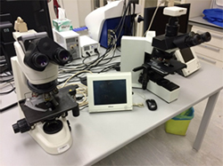
| Nikon E90i Fully Motorised Upright Fluorescence This microscope is equipped with blue, green and red filters for imaging common fluorescent dyes in these ranges. |
 | Nikon TE2000U Inverted Fluorescence (in School of Medicine’s Culture Lab) |
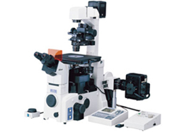
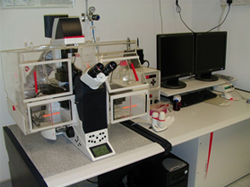
| Leica AF6000LX Live Cell Imaging
|
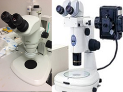
| Nikon Stereoscopic microscope (SMZ1000, SMZ1500)
|
| Zeiss AxioZoom Fluorescence StereoZoom (in Animal Research Platform)
|
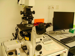
| Confocal Microscope | |
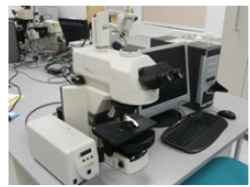
| Nikon C1si Inverted Confocal Laser Microscope
|
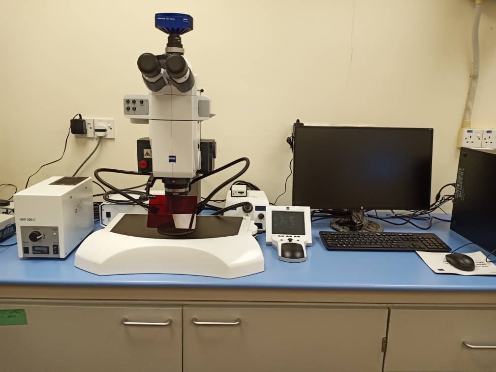
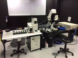
| Multiphoton Microscope | |
| Leica SP8 Multiphoton microscope
|
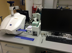
| Automated Slide Scanning | |
| Zeiss Mirax Midi
|
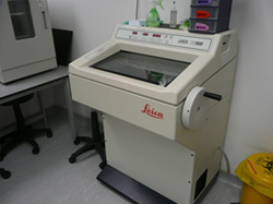
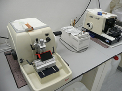
| Histology Lab Equipment | |
| Leica CM1860 Cryostat
|
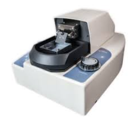
| Leica RM2035 Microtome
|
 | D.S.K Microslicer ZERO 1N Vibratome
|
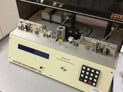
| Micropipette Puller
|
Specialist Services
Our team provides advanced microscopy instrumentation and analytical techniques to a large research community. Ranging in complexity from the cytology, histology samples to live cell or in vivo imaging, we will guide and train users to perform experiments, produce high-quality images and extract analytical data.
Specialist service #1: Advanced light and fluorescence microscopy
Our instrumentation provides a solid platform of advanced light and fluorescence microscopy techniques, including, time-lapse, slide scanning (in conjunction with our histology facilities) and image tiling, and live-cell imaging on slides, chambers or microplates. Both upright and inverted microscopes are available, and all systems are supported by a comprehensive range of professional software for bioimage analysis to provide quantitative results.
Specialist service #2: Live-cell imaging is one of our specialties
Live-cell imaging instrument is equipped with a temperature-, humidity- and CO2 -levels controlled chamber and deconvolution function , which supports live and long term imaging experiments.
Specialist service #3: Optical sectioning and 3D analysis
Our range of instrument modalities includes confocal laser scanning (CLSM) and multiphoton microscopes (MP). Imaging deeper tissue can be done by multiphoton imaging in live, fixed or cleared tissue microscopy which is capable of imaging to a depth of ~800 μm with specialised objectives.
Specialist service #4: Image analytics
Extracting and understanding bioimaging data is crucial, and handling big datasets is often a bottleneck in research. Our staff are available to train scientists and students in the analytical software with the help of Monash Micro Imaging at Monash Australia.
Other capabilities
Histology-specialised equipment includes cryostats, microtomes, vibratome and laser-capture microdissection for histological and morphological analysis. An automated brightfield and fluorescent slide scanner is also available for digitalization of histology or cytology specimens direct to your screen.
Download eFlyer here.
Access and Training
The Optical Imaging Platform is open to all members of the University, external academic institutions and industry.
In house training is provided.
Request Service/ Booking
Request for services on Optical Imaging Platform iLab
Publications and Acknowledgements
Your use of the Monash Malaysia Research Infrastructure is heavily subsidised by the University. To comply with internal reporting obligations, the platform must report on the number and type of publications produced by users of the platform/ facility. It is essential that we have this information so that we can continue to provide the advanced instrumentation, staff, training and assistance that you and other researchers require to undertake your work.
It is a condition of use of platform that:
1. You acknowledge the platform and any significant assistance provided by the staff members in your publications;
2. Platform staff members who made a significant scholarly contribution should be co-authors on the publication, in accordance with Monash’s Research Authorship And Attribution Policy, see Research Authorship and Attribution Policy.
Please acknowledge MUMOIP in your publications by including:
"The authors acknowledge the use of instruments and scientific and technical assistance at the Monash Malaysia Optical Imaging Platform (MUMOIP)”.
Or,
if the work was carried out with help from MUMOIP staff (but not enough to justify coauthorship):
"The authors acknowledge the use of instruments and scientific and technical assistance of Name of OIP Staff Member at the Monash Malaysia Optical Imaging Platform (MUMOIP).”
Please always notify the relevant platform when acknowledgment is made.
Location
Building 3, Level 3,
Jeffrey Cheah School of Medicine and Health Sciences,
Monash University Malaysia.